New Insights into Visual Processing in the Mouse Brain
Understanding How Visual Information is Processed in the Brain
At a Glance: Scientists have successfully mapped the intricate connections between hundreds of thousands of neurons within a minuscule region of the mouse brain, while also examining their firing patterns in response to various visual stimuli. This groundbreaking research sheds new light on the brain's mechanisms for processing visual information, ultimately helping to reconstruct the images that humans and other mammals perceive.
The Allen Institute, a leader in brain research, emphasizes that understanding information processing in the human brain involves the electrical firing of approximately 86 billion neurons, which establish trillions of connections among themselves. The secrets behind how the brain enables us to think, feel, and act are embedded within the complexity of these neural pathways and the rapid electrical signals that traverse them in mere milliseconds.
Recent advances in mapping neural connections, supported by the National Institutes of Health's (NIH) Brain Research Through Advancing Innovative Neurotechnologies Initiative, commonly known as the BRAIN Initiative, have made significant progress in unveiling the mysteries of the brain. This initiative is particularly focused on mapping the brains of various species, including mice, fruit flies, and humans.
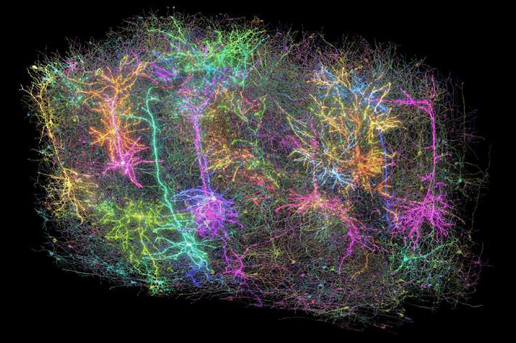
In what marks a remarkable achievement, researchers from the Machine Intelligence from Cortical Networks (MICrONS) Consortium have mapped connections among hundreds of thousands of cells in a tiny region on the surface of the mouse brain that is crucial for visual processing. This analyzed area measures just one cubic millimeter, roughly comparable to the size of a grain of sand. The researchers superimposed their neuronal map with firing patterns recorded in response to visual stimuli. Their findings were published in a collection of eight papers in the esteemed journals Nature and Nature Methods on April 9, 2025.
The experimental process began with the scientists presenting video clips to a mouse whose neurons emitted light whenever they fired. This innovative technique allowed the research team to meticulously record the firing patterns of nearly 76,000 neurons during the viewing sessions.
To create a detailed mapping of neuronal connections, the researchers meticulously cut nearly 28,000 ultra-thin slices of brain tissue, which they then imaged using advanced electron microscopy. The daunting task of reconciling these images into a cohesive whole involved painstakingly stitching them together to accurately align the connections throughout the entire volume. This process was further enhanced over several months through deep learning artificial intelligence (AI) algorithms, followed by rigorous manual and automated proofreading to ensure accuracy.
Remarkably, even in this minuscule area of the brain, the scientists discovered approximately four kilometers of axons, which are the long fibers that enable nerve cells to communicate with one another. These axons are intricately intertwined, forming over half a billion synaptic connections across more than 200,000 cells. The cataloged cells included various types of neurons as well as a range of support cells, highlighting the complexity and diversity of brain architecture. The team demonstrated that the connectivity patterns could be instrumental in identifying cell types that might be challenging to differentiate based solely on their shapes.
The researchers constructed and validated deep learning predictive models that correlate neuronal activity with physical connectivity. As a result, this mapping represents the most comprehensive data set ever assembled that links brain structure to neuron function in an active mammal. This profound insight offers new perspectives on how the brain processes visual stimuli. Notably, the foundational architecture of the brain cortex remains remarkably consistent across different mammalian species, from mice to humans.
Dr. John Ngai, director of the BRAIN Initiative, elaborates, While the current findings focus on a tiny fraction of the brain, they reveal the complex connections between the cells and show how those connections are wired to produce functional responses. This information, which was previously beyond our reach, could help us understand how the brain functions normally and offer insights into what goes awry as a result of various disorders or injuries.